Unfolding the Riddles of Proteins
RELATED LINKS
Look in on Baylor biochemist Bryan Shaw and one of the first things you'll notice on his office shelf are several objects that resemble the ball of ice atop a snow cone, only with some of it melted away. They are 3D models of proteins that, on brief examination, display a simple form that belies the unimaginably complex structure folded up within — a complexity that even the most powerful supercomputers have yet to fully fathom.
"I love proteins," Shaw confesses. "I really like the way they look. A lot of people are in biochemistry because they saw these beautiful, elegant protein structures and they got sucked in, they wanted to know more about it."
It is a fascination that began when he was an undergraduate at Washington State.
"We used a textbook written by a wonderful professor named Joan Valentine. She was at UCLA and I was interested in her work. My undergraduate professor said, 'Why don't you apply,' so I did. She had a big influence on me."
At the time, proteomics — the large-scale study of protein structure and function — was exploding as a relatively young branch of biochemistry. Valentine introduced him to metalloproteins, proteins that have metal ions bound up in their structures. It's estimated that over a third of all proteins are metalloproteins, yet for all their prevalence, some of their chemical properties weren't yet fully characterized.
He departed UCLA with Ph.D. in-hand, having landed a plum fellowship in the laboratory of legendary Harvard chemist, Dr. George Whitesides. It was there Shaw began to acquire an appreciation for the role that a protein's net electrostatic charge might play in the way it interacts with nearby proteins, especially those that clump together into masses that can become toxic to brain cells.
It's these protein aggregates that are thought to be a major factor in the development of ALS — amyotrophic lateral sclerosis — in Alzheimer's and in several lesser-known but still devastating neurological disorders. Shaw finds the connection between electrostatic charge and aggregation to be convincing.
"The connection appears to be very, very strong," he says. "With many protein aggregation diseases like Lou Gehrig's disease, most of the cases are sporadic – there's no known cause. But a small percentage are inherited, and when we look at how the mutations in these genes affect the proteins that prove to be toxic, we're finding that a lot of them promote aggregation by destabilizing the protein or altering its structure. In fact, most mutations do that, but some of them do nothing but lower the net charge. That's the only thing we've found that could be making these proteins aggregate more rapidly."
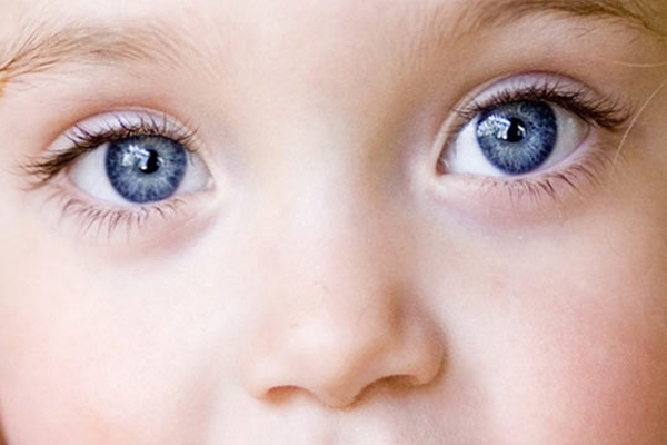
Shaw's Harvard research was advancing steadily when, in May 2008, family circumstances intervened. His wife Elizabeth discovered that in some of the flash photos of their three-month-old son Noah, his right eye gave off a white reflection, unlike the red reflection on his left eye.
A trip to the pediatrician and then to an ophthalmologist yielded devastating news: Noah had retinoblastoma, a serious, sometimes fatal form of eye cancer. Months of anguished trips to clinics for evaluation and treatment passed, ultimately resulting in the loss of one of Noah's eyes.
Working with Harvard doctors who were treating Noah, Shaw digitized stacks of photos of Noah and other retinoblastoma patients to determine just how early photographs may reveal a tumor. They learned that photographs could show the presence of a tumor very early indeed. One photo of Noah taken when he was only 12 days old displayed the tumor at a time when it was much smaller and might have required less extensive treatment. The finding is getting widespread exposure in both the medical and popular media.
Happily, today Noah is an energetic, curious kid with a promising future ahead of him. And while the experience did not diminish his enthusiasm for protein research, it left Shaw deeply sensitive toward visually disabled children. That sensitivity imparts a new spin to his work at Baylor — and also helps explain the snow-cone-like models in his office.
"It turns out that in chemistry, the most underrepresented group of students are blind students," Shaw says. "And the reason of course is the preponderance of visual tools. We look at protein structures on a computer screen with molecular graphic simulations. How are we going to show that to a student who's blind?"
The desire to convey the intricate complexity of protein structures to blind students led him to merge the vast information resources of the proteomic database with the now-burgeoning technology of three-dimensional printing.
"We can go into the protein databank and get the coordinates of every atom of a protein as measured by X-ray crystallography. Then we can feed that into a 3D printer and print up highly accurate models of protein molecules like the ones you see over there," he says, pointing to the models. "We have to harness every remaining sense they have — their fingertips, their nose, their mouth, their hearing, whatever!" That's why some of his models are smaller and mounted on a stick like a lollipop. Those allow blind students to explore protein structure with the highly tactile lips and tongue.
"Educating people is important, but inspiring them is more important," Shaw declares. "Once you inspire people, it becomes a lot easier to educate them for the rest of their lives. They might not be able to understand the complexity of something, but if it's fascinating and it's beautiful and if you can inspire them, they're hooked."
The man speaks from experience.
Dr. Shaw's work is funded in part through grants from the National Science Foundation and the U.S. Department of Defense. He also recently received a five-year, $405,000 NSF CAREER award, and a grant from the Welch Foundation. More information on Dr. Shaw's research can be found here.